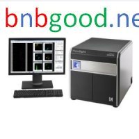
FlowSight Quantitative Imaging Analysis Flow Cytometer, Cell Biology Instrument
Inhouse product
-

LUB12 standard controll unit
$60.00 -
Barbed Wire
$0.00 -
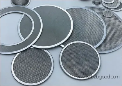
Perforated Sheet Mesh
$0.00

Life science research has advanced into the field of cellular studies. Scientists are eager to explore the deep and comprehensive information of cells. They not only provide information about cell populations but also focus on the differences between individual cells. They can not only observe the characteristics of cell phenotypes but also study the functions of cells. However, current flow cytometry techniques are not yet sufficient to meet the application needs of these scientists. The FlowSight Multi-dimensional panoramic flow cytometry, launched by Merck Millipore in 2012, for the first time enabled comprehensive cell analysis from groups to individuals, from phenotypes to functions, leading flow cytometry into a new era.FlowSightIt is the next generation of expert-level flow cytometry.
FlowSight is a compact yet powerful next-generation expert-level flow cytometer. The system is designed to revolutionize the improvement of the signal-to-noise ratio and the sensitivity of fluorescence detection. In addition to the traditional fluorescence intensity information, 12 detection channels can be calibrated to obtain bright field, dark field, and 10 fluorescence images of each cell. FlowSight can be equipped with up to four laser tubes (405, 488, 561, 642 nm) and a 785 nm laser forSSC信号检测。该设备还可以配备96孔板自动采样系统(AutoSampler)。FlowSight具有低于10 MESF的高检测灵敏度,特别是在弱信号检测方面,具有明显的优势。
FlowSightBreaking through the limitation of traditional flow pattern and realizing circle door no longer depends on guessing
FlowSight differs from other traditional flow cytometry methods because it can display the true image of each cell. For each cell, it can generate 12 images. These images can identify fluorescence from the cytoplasm, cell membrane, and nucleus. By clicking on each point in the scatter plot or histogram, users can view the corresponding cell image. As a result, users no longer need to guess whether the data is accurate. In FlowSight, once the data is set up, users can confirm the accuracy of the data. The accuracy of the data can be verified by observing the cell images inside and outside the "door."
Contact Information:
17338135972
010-86468961-808
E-mail: niebaojin@gotobio.com
Homepage: www.gotobio.com
Related products
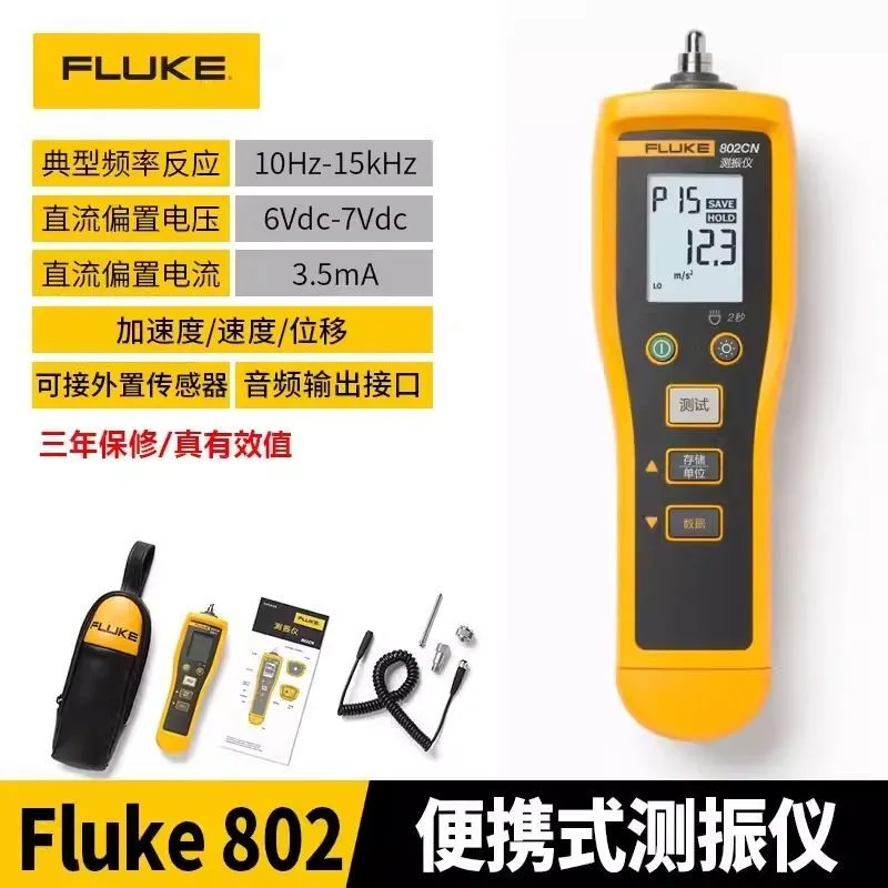
Fluke-805

Folding arm crane
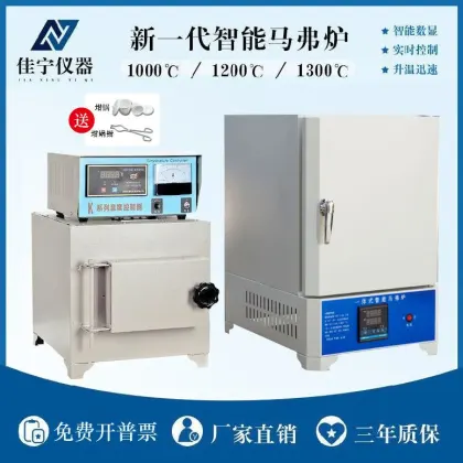
SX2智能马弗炉热处理灰分退火淬火炉高温箱式电阻炉实验电炉工业
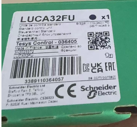
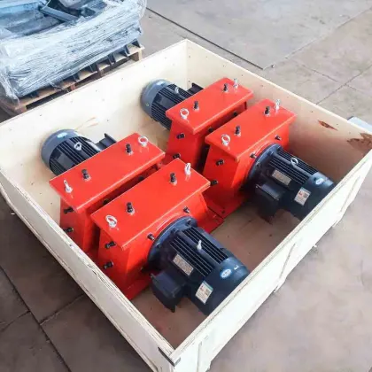
LUB12 standard controll unit

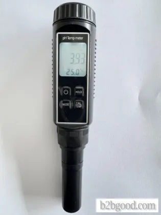
PH- A6 防水笔式高精度 pH / 温度计

Contact Info
- Address: Dezhou city,Shandong,China
- Phone: +86-15166945837
- Email: dzheibei@sohu.com